Special Testing
Beyond the Eye Chart: Advanced Testing for Clearer Insights
At Concord Eye Care Center, we believe that true eye care extends far beyond the traditional eye chart. Our commitment to your ocular health drives us to employ cutting-edge diagnostic technologies that provide a comprehensive view of your eyes’ structure and function. These advanced tests allow us to detect potential issues at their earliest stages, often before you experience any noticeable symptoms.
Our suite of specialized equipment enables us to examine your eyes with unprecedented detail and accuracy. From mapping the intricate landscape of your retina to assessing your peripheral vision and measuring intraocular pressure without the need for eye drops, these non-invasive procedures offer invaluable insights into your overall eye health. By utilizing these advanced diagnostic tools, we can tailor our care to your unique visual needs, ensuring that you receive the most precise and effective treatment possible.
Our special tests allow us to detect and monitor eye conditions with unprecedented accuracy and comfort. Let’s explore three of our advanced diagnostic tools:
Optomap® Retinal Exam
The Optomap® is a revolutionary diagnostic tool that allows us to capture a detailed image of your retina – the back of your eye – in a single, quick scan.
How it works:
- You simply look into the device one eye at a time
- A comfortable, low-intensity laser scans your retina
- In less than half a second, we have a comprehensive, digital image of your retina
Benefits:
- Non-invasive and painless
- No eye dilation required in most cases
- Allows us to detect issues like retinal detachment, glaucoma, and even signs of diabetes or high blood pressure
- We can compare images year-over-year to track changes in your eye health
What to expect during your optomap® exam:
When you come in for your Optomap® exam, you’ll be seated comfortably in front of the machine. Our trained technician will guide you through the process, which typically takes only a few minutes. You’ll look into the device one eye at a time, and you might see a flash of light as the image is taken. There’s no discomfort, and the results are available immediately for our optometrists to review with you.
Why we use optomap® at Concord Eye Care Center:
We’ve chosen to invest in Optomap® technology because it allows us to provide a higher standard of care. Traditional methods of examining the retina can be time-consuming and uncomfortable, often requiring dilating eye drops. With Optomap®, we can perform a thorough retinal exam quickly and comfortably, allowing us to detect and monitor a wide range of eye conditions more effectively.
Zeiss / Humphrey Visual Field Testing
Visual field testing helps us measure the entire scope of your vision, including your peripheral (side) vision.
How it works:
- You’ll sit in front of a bowl-shaped instrument
- Focusing on a central point, you’ll indicate when you see lights flash in your peripheral vision
- The test typically takes 5-10 minutes per eye
Benefits:
- Helps detect vision loss from glaucoma, stroke, and other neurological issues
- Allows us to monitor progression of known conditions
- Essential for assessing your ability to safely perform tasks like driving
Preparing for your Visual Field Test:
Before your appointment, make sure you’re well-rested, as fatigue can affect the test results. If you wear glasses or contact lenses, bring them with you. During the test, you’ll need to concentrate, so we recommend avoiding caffeine if it makes you jittery. The test can be repeated if necessary, so don’t worry if you think you’ve made a mistake.
Interpreting your Visual Field Test results:
After the test, our optometrists will carefully analyze the results. We’ll look for any areas of reduced sensitivity in your visual field, which could indicate various eye or neurological conditions. We’ll explain the results to you in detail, showing you the visual field map and discussing any findings. If we detect any issues, we’ll work with you to develop an appropriate treatment or management plan.
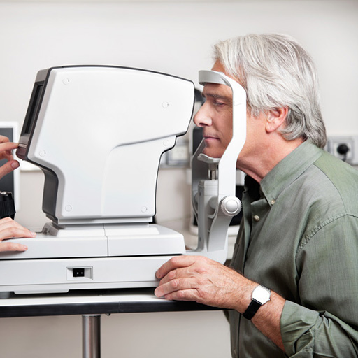
iCare Tonometry
The iCare tonometer is an advanced tool we use to measure the pressure inside your eye, which is crucial for detecting glaucoma early. Unlike traditional methods, it uses a tiny probe that gently touches your eye for just a millisecond. It’s so quick and comfortable that most patients don’t even feel it. This allows us to get accurate readings easily, even for sensitive patients or children. By using this technology, we can detect potential issues earlier and monitor your eye health more effectively, helping to preserve your vision long-term.
How it works:
- A small, lightweight probe gently touches your cornea for a split second
- The device measures the pressure inside your eye
- The process is so quick and gentle, most patients don’t even feel it
Benefits:
- No eye drops or air puffs needed
- Extremely accurate readings
- Comfortable and stress-free for patients of all ages
- Allows for frequent pressure checks when monitoring glaucoma
The iCare experience at Concord Eye Care Center:
When you come in for an iCare tonometry test, you’ll be comfortably seated while our technician explains the process. The test is so quick and gentle that many patients are surprised when it’s already over. Unlike traditional “air puff” tests, which can be startling, iCare tonometry is virtually unnoticeable. This makes it an excellent option for patients of all ages, including children and those with anxiety about eye exams.
Why accurate pressure readings matter:
Intraocular pressure is a crucial measurement in eye health, particularly in the detection and management of glaucoma. By using iCare tonometry, we can obtain highly accurate pressure readings without the need for numbing eye drops or air puffs. This allows us to perform the test more frequently if needed, which is especially valuable when monitoring glaucoma or ocular hypertension. Regular, accurate pressure readings help us ensure that your treatment is effective and make timely adjustments if necessary.
At Concord Eye Care Center, we believe in combining our clinical expertise with cutting-edge technology to provide you with the best possible care. These special tests allow us to detect potential issues early, monitor existing conditions more effectively, and ensure your eyes stay healthy for years to come.
Have questions about these tests or want to schedule an appointment? Don’t hesitate to contact us. Your vision is our priority, and we’re here to help you see clearly and comfortably.
OPTOS Retinal Exam
Annual eye exams are vital to maintaining your vision and overall health. We offer the optomap® Retinal Exam as an important part of our eye exams. The optomap® Retinal Exam produces an image that is as unique as you fingerprint and provides us with a wide view to look at the health of your retina. The retina is the part of your eye that captures the image of what you are looking at, similar to film in a camera.
Many eye problems can develop without you knowing. You may not even notice any change in your sight. But, diseases such as macular degeneration, glaucoma, retinal tears or detachments, and other health problems such as diabetes and high blood pressure can be seen with a thorough exam of the retina.
An optomap® Retinal Exam provides:
- A scan to show a healthy eye or detect disease.
- A view of the retina, giving your doctor a more detailed view than he/she can get by other means.
- The opportunity for you to view and discuss the optomap® image of your eye with your doctor at the time of your exam.
- A permanent record for your file, which allows us to view your images each year to look for changes.
The optomap® Retinal Exam is fast, easy, and comfortable for all ages. To have the exam, you simply look into the device one eye at a time and you will see a comfortable flash of light to let you know the image of your retina has been taken. The optomap® image is shown immediately on a computer screen so we can review it with you
ZEISS / Humphrey Visual Field Testing
Visual Field testing can help save vision because it is another test used to diagnose or rule out glaucoma and other neurological disorders that affect vision. This simple, but effective service has saved lives by detecting various medical conditions such as strokes, brain tumors, and other neurological defects.
Icare Tonometry
The Icare tonometer is based on a proven accurate measuring principle, in which a very light probe is used to make momentary and gentle contact with the cornea. The measurement is barely noticed by the patient and often does not even cause corneal reflex. The device not only makes IOP measuring a more pleasant experience on all patients, it is also an important break-through for succeeding with non-compliant patients (f.e. children and dementia patients).
Optical Coherence Tomographer (OCT)
Our OCT helps us better manage glaucoma and diseases of the retina because this technology allows the eye doctor to see the deep tissue layers in the eye. Similar to ultrasound, this diagnostic technique employs light rather than sound waves to achieve higher resolution pictures of the structural layers of the back of the eye. These high-definition images are the only way that they can actually see beneath the surface to the nerve fiber layers where damage occurs. Up until now, eye doctors had to use other tests to indicate damage in this critical area of sight. Common eye diseases such macular degeneration, diabetic retinopathy, and glaucoma are detected early by the OCT when the diseases can be more effectively treated.
Corneal Topography
Corneal topography is a computer assisted diagnostic tool that creates a three-dimensional map of the surface curvature of the cornea. The three-dimensional map is a valuable aid to the examining optometrist and can assist in the diagnosis and treatment of a number of conditions; in planning cataract surgery and intraocular lens (IOL) implantation (plano or toric IOLs); in planning refractive surgery such as LASIK, and evaluating its results; or in assessing the fit of contact lenses.
i.Terminal 2
Lens fitting plays a key role in maximizing visual comfort, as fitting errors can cause up to a 40% loss in lens performance. i.Terminal 2 captures and calculates your patient´s individual parameters with the click of a button and a precision of 0.1 mm which can result in a decreased complaint rate, reduced non-adapts and more relaxed vision for your patients.
i.Scription
i.Profiler® plus not only provides you a better prescription, it gives you access to an optimized individual lens solution with i.Scription technology for improved color and contrast vision as well as improved night vision. i.Scription involves an innovative patented algorithm which combines the subjective refraction values with the i.Profiler plus and ocular wavefront aberrometry data to calculate an individualized prescription to 1/100th of a diopter – especially for lower light situations.
Retinal Digital Imaging – Fundus Photography
A high-definition digital image of the retinal area helps your eye doctor diagnose and manage eye diseases in the delicate retinal area. Damage to these delicate structures of the retinal area is often the first sign of systemic diseases such as MS, diabetes and more. The retina is the “window to the body” and routine retinal imaging can help your eye doctor monitor the changes in your eye health from year to year.
Glaucoma Diagnosis (GDx)
Our GDx helps us better manage glaucoma and diseases of the retina because this technology allows the eye doctor to see the deep tissue layers in the eye. These images are the only way that they can actually see beneath the surface to the nerve fiber layers where damage occurs. Up until now, eye doctors had to use other tests to indicate damage in this critical area of sight. Glaucoma can be detected early by the GDx when the diseases can be more effectively treated.